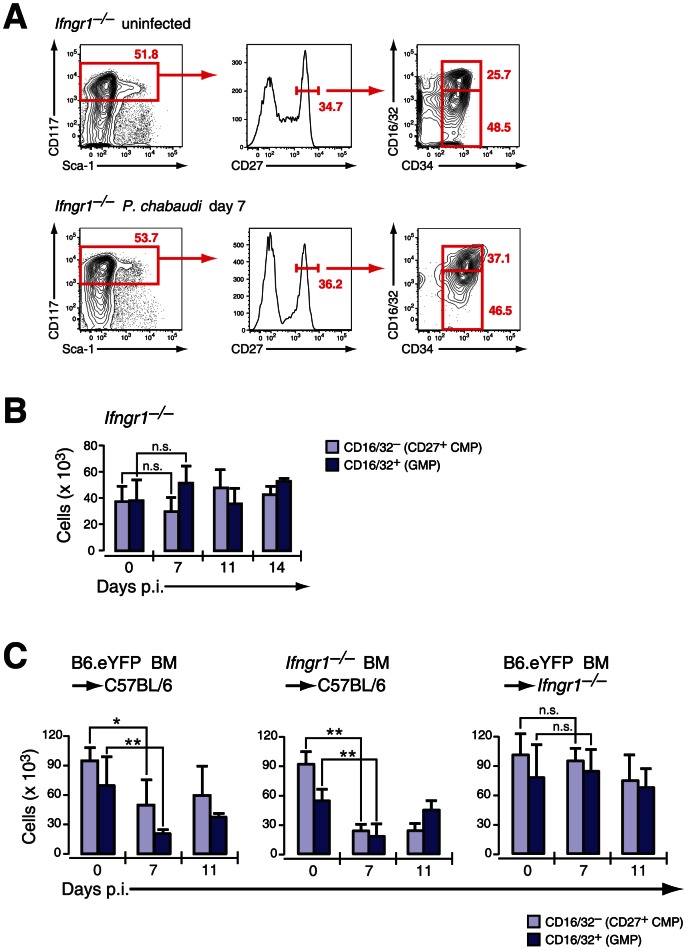
Figure 4. Infection-induced decrease of early myeloid progenitors is critically dependent on IFN-γ signaling.
A. Phenotype of LIN− BM cells in Ifngr1-null mice uninfected and at day 7 after infection with P. chabaudi stained for indicated marker. Representative FACS plots of four experiments with each 5 mice per group are shown. B. Absolute numbers (per femur/pair) for Ifngr1-null LIN− c-Kithi CD27+ cells are shown as mean ± SEM per individual subsets obtained form three experiments with 5–6 mice per group (n.s.: P>0.05, Mann-Whitney U-test). C. Absolute numbers of myeloid progenitors in radiation chimeras (Hosts: C57BL/6, left and middle; Ifngr1-null, right chart) reconstituted either with B6.Rosa26eYFP or Ifngr1-null and 8–9 weeks after transplantation infected with P. chabaudi. Two different sets of chimeras were infected and 5–9 animals per time point each were analyzed. Data represent mean ± SEM of cell number per femur/pair (*: P≤0.05; **: P≤0.01, Mann-Whitney U-test).

