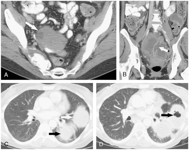
FIGURE 2.
A, Axial contrast-enhanced CT image in a 50-year-old woman with metastatic melanoma showing a pelvic tumor deposit (arrow). B, Coronal reformatted contrast-enhanced CT image after treatment with 2 cycles of XL184 shows a wide-mouth fistula (arrow) between a loop of small bowel in the pelvis and the tumor mass, which is now largely cystic and fluid-filled. C, Axial contrast-enhanced CT image obtained at the same time as A shows 2 pulmonary metastases (arrows) in the left lower lobe. D, Axial contrast-enhanced CT image obtained at the same time as B shows new air foci (arrow) in the anterior metastasis, consistent with development of a fistula between the tumor and the tracheobronchial tree.

