
Figure 2.
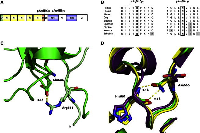
PDGFR-β Substitutions in Familial Infantile Myofibromatosis
(A) Domain structure of PDGFR-β and position of identified mutations. Abbreviations are as follows: SP, signal peptide; Ig, immunoglobulin-like C2 domain; TM, transmembrane domain; JM, juxtamembrane domain; KD1, first split kinase domain; KI, kinase insert; KD2, second split kinase domain; CT, C-terminal tail domain.
(B) Amino acid conservation across species for the mutated residues.
(C) A ribbon diagram showing the interaction between Arg561 and Glu644 in the PDGFR-β model. The backbone of the model is shown in green, with residues of the JM domain colored light green. The p.Arg561Cys substitution would abrogate this interaction.
(D) A ribbon diagram showing the interaction between Asn666 and His661 in the PDGFR-β model (green). The structure of the autoinhibited (yellow) and active (purple) forms of KIT kinase are shown for comparison. A p.Asn666Lys change would abolish the interaction linking Asn666 and His661.