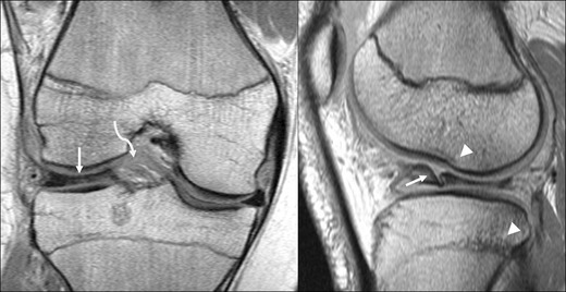
Fig. 12.

Discoid lateral meniscus. Coronal (left) and sagittal (right) PD images in a 16-year-old boy, showing a bulky discoid lateral meniscus (arrows) with a flap tear, as well as a ruptured ACL (curved arrow), femoral condylar notch impaction fracture and tibial contusion (arrowheads)
