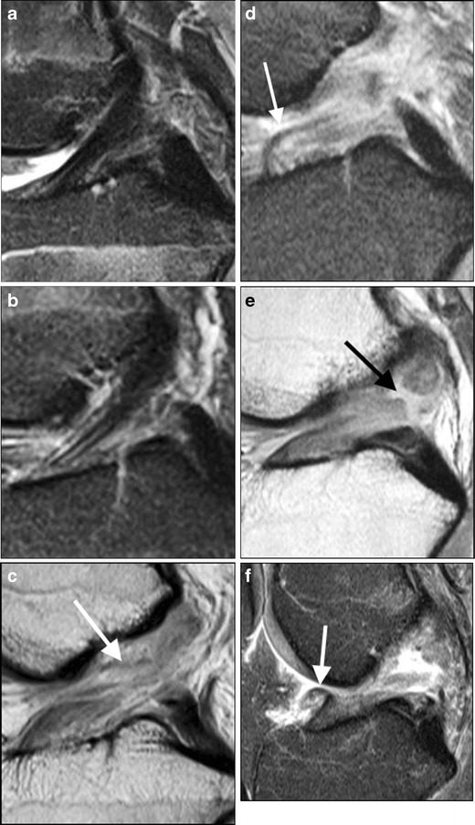
Fig. 2.

Spectrum of ACL injury in sagittal plane (a–f). Proton-density (PD) images (with fat saturation [FS] except c and e) in paediatric patients age 13–16 years, showing (a) intact ACL, b intact but somewhat thin ACL, (c) our only surgically confirmed high grade partial tear of ACL, demonstrating lax fibres (arrow), (d) full thickness tear at midsubstance with some intact fibres near tibial attachment (arrow), (e) obvious full thickness tear (arrow), (f) full thickness tear with anteriorly flipped distal ligament fibres (arrow) and anterior tibial translation
