Fig. 4.

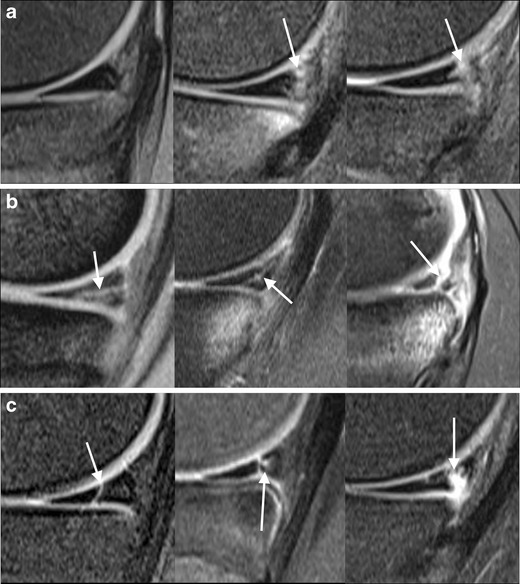
Spectrum of posteromedial corner injury on sagittal T2 FS images (image d is T2 GRE). a Intact meniscus: normal (left); increased signal at meniscocapsular junction representing minor strain, not meeting criteria for tear (middle; note the underlying tibial contusion); meniscocapsular junction tear, with no dark meniscal tissue posterior to the high-signal cleft (right). b Meniscal tears: horizontal extending to undersurface (left); oblique tear with tibial contusion (middle); displaced oblique tear with tibial contusion (right). c Meniscal tears: vertical tear (left); vertical and intrasubstance tear (middle); displaced complex tear, with vertical and horizontal components (right)
