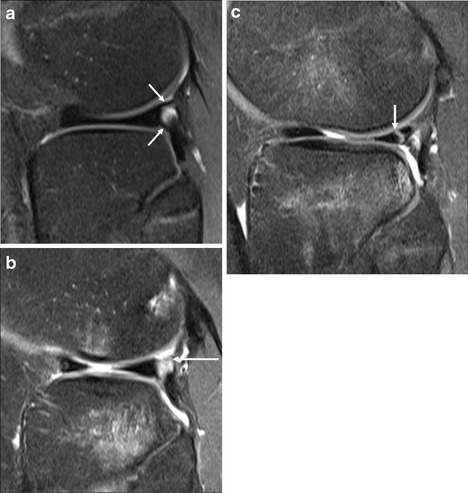
Fig. 6.

Posterolateral corner on sagittal PD FS images. a Normal, with intact lateral meniscus, and superior and inferior popliteomeniscal struts (arrows). b Tear of the superior (arrow) and inferior popliteomeniscal struts, with intact meniscus but associated tibial and femoral marrow contusions. c Vertical tear of lateral meniscus (arrow), with irregular appearance of inferior popliteomeniscal strut suggesting incomplete tearing, marrow contusions similar in pattern to b, and a partly visualised lateral femoral condyle impaction fracture
