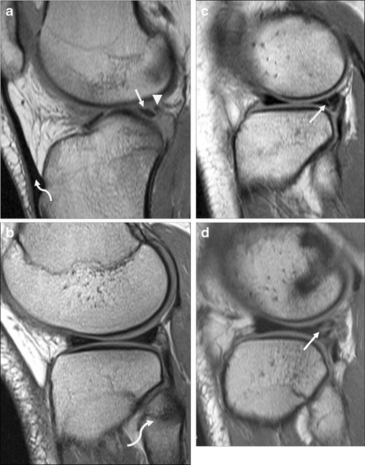
Fig. 7.

Posterior lateral meniscus tear. a Sagittal PD image showing cleft between posterior third of lateral meniscus (arrow) and the meniscofemoral ligament of Wrisberg (arrowhead). These structures are normally separate medially, near the intercondylar notch (note that this slice contains the patellar tendon, curved arrow), but merge laterally. b Normal appearance on a more lateral slice, where fibular head is visible (curved arrow): the posterior lateral meniscus is a single triangular structure. c Oblique, mainly vertical undisplaced tear of the posterior lateral meniscus (arrow). The cleft is abnormal when present laterally on an image where the fibular head is visible. Note that Fig. 6c shows a displaced similar tear. d Complex tear of the same region of meniscus (arrow)
