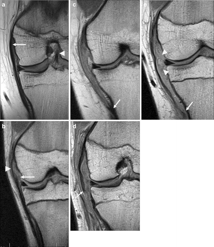
Fig. 9.

Medial collateral ligament on coronal PD images, reoriented as if all were left knees for comparative purposes. a Normal MCL, appearing slightly thicker proximally (arrow) than distally, which is typical. Also note the intact ACL (arrowhead). b A low-grade partial tear of deep MCL fibres including medial meniscofemoral ligament (arrow). Superficial fibres are intact (arrowhead). c High-grade sprain involving nearly the entire length of MCL; note normal appearing tibial attachment (arrow). d MCL rupture just distal to knee joint line (arrow). e A distal rupture near tibial attachment (arrow), with associated tears of deep fibres including meniscofemoral and meniscotibial ligaments (arrowheads)
