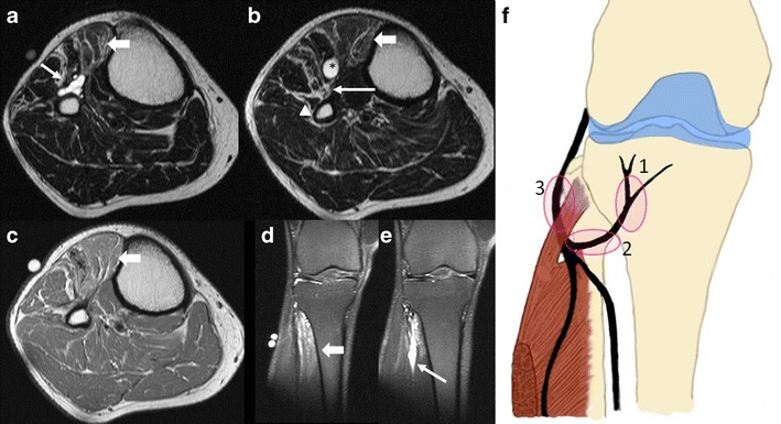
Fig. 11.

Recurrent intraneural ganglion. Axial T2-WI at the level of the right fibular neck (a) shows the horizontal part of the recurrent articular branch at the 12 o’clock position. The level just below (b) shows extension of the cyst in close relationship with the deep peroneal nerve (arrow), situated between the cyst and the fibular neck. More laterally the superficial peroneal nerve can be discerned (arrowhead). On both images the signal intensity of the anterior tibial muscle (thick arrow) is slightly higher when compared to the surrounding muscles. Axial T1-WI (c) shows no significant fatty infiltration in the anterior compartment. Coronal fat-suppressed T2 PD-WI (d) shows a hyperintense signal in the most proximal part of the anterior tibial muscle (thick arrow), in keeping with denervation oedema. The second, more posterior coronal fat-suppressed T2 PD-WI (e) shows the hyperintense ganglion (thin arrow) with a slightly beaded appearance along the course of the deep peroneal nerve. Irritation of the peroneal nerve by the ganglion was confirmed on the EMG. The ganglion was surgically drained and curettage of the proximal tibiofibular joint was performed. Schematic drawing (f) showing the sites where the typical signs of an intraneural ganglion can be observed. The ascending part of the articular branch is the location of the tail sign (1); the transverse limb sign can be observed on the horizontal part of the articular branch (2). When the intraneural ganglion extends proximally in the common peroneal nerve a signet ring sign can be observed (3)
