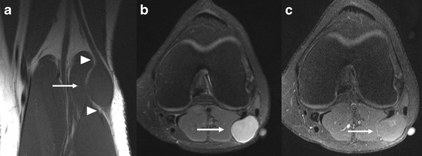
Fig. 12.

a-c Schwannoma of the left common peroneal nerve. Coronal T1-WI (a) showing a spindle shaped lesion (arrow) in the common peroneal nerve at the level of the lateral femoral condyle, with a signal that is isointense to the surrounding muscle. The peroneal nerve is seen entering proximally and exiting distally (arrowheads). A split-fat sign is visualised around the lesion. Axial fat-suppressed T2-WI (b) shows a homogeneous hyperintense signal (arrow). Axial fat-suppressed T1-WI after IV administration of gadolinium contrast (c) shows almost no enhancement (arrow)
