
Fig. 14.
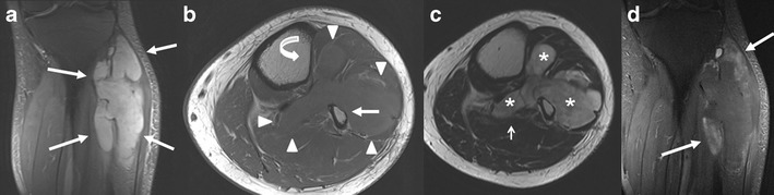
Myxoid liposarcoma compressing the peroneal nerve. Because the patient had café au lait spots, a diagnosis of a malignant PNST in a patient with NF1 was initially proposed, with the request for pathological confirmation. Coronal fat-suppressed PD T2-WI (a) shows a polylobulated homogenously hyperintense mass (thin arrows) mostly located in the lateral compartment of the left lower leg, larger than 5 cm. Axial T1-WI (b) shows the mass (arrowheads) extending in the anterior, lateral and deep posterior compartment. Note scalloping on the anterior side of the fibula (thick arrow) and the lateral side of the tibia (curved arrow). Axial T2-WI (c) shows a heterogeneous mostly hyperintense signal in the mass (asterisk), clearly delineating it from the surrounding muscle. There is no fat plane between the soleus muscle and the superficial posterior compartment (arrow), strongly suggesting extension in all four compartments of the lower leg. No muscle oedema is present. Coronal fat-suppressed T1-WI after IV administration of gadolinium contrast (d) shows inhomogeneous peripheral enhancement of the mass (arrows)