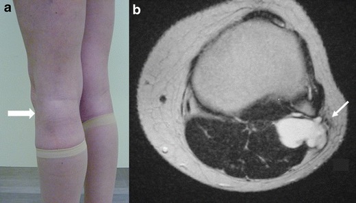
Fig. 16.

Extraneural ganglion. Photograph of the left knee (a) shows a focal swelling on the posterolateral corner of the knee (thick arrow). Axial T2-WI (b) shows a cyst posterior to the proximal tibiofibular joint, displacing the common peroneal nerve laterally (arrow)
