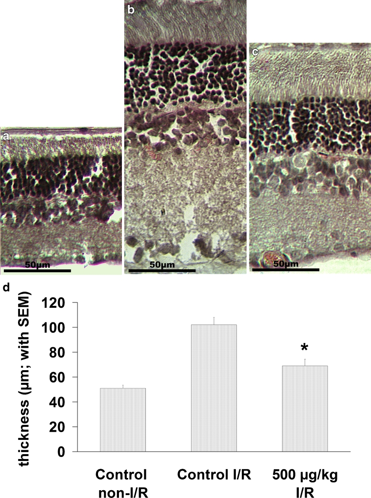
Fig. 3.
Effect of α-MSH on I/R-induced retinal edema (group I-b). Sagittal sections of the rat retina showing the structure of layers in nonischemic retinal tissue of vehicle-treated (control) animals (a), I/R-injured retinal tissue from vehicle-treated (control) rats (b), and I/R-injured retinal tissue from animals treated with 500 μg/kg α-MSH (c). d The effect of α-MSH on the extent of retinal edema following I/R injury, in the inner plexiform layer. Thickness was measured using a manual scale on each glass slides. Mean ± SEM; *p < 0.05 vs. control I/R

