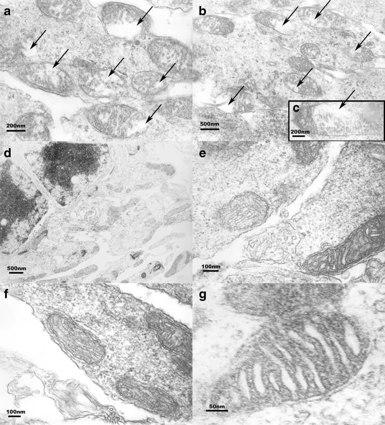
Fig. 4.
EM studies (group I-b). a–c Mitochondria in I/R-injured inner retinal cells from rats not treated with α-MSH. These images demonstrate disintegration of the mitochondria due to formation of interior vacuoles (arrows). Mitochondria in I/R-injured inner retinal cells from animals treated with 500 μg/kg α-MSH are shown in (d–g). No vacuolization is seen here

