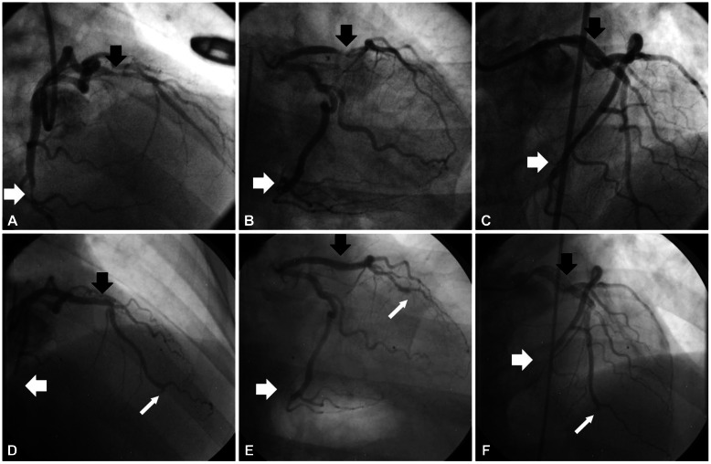
Fig. 1.
At the first admission, left coronary artery angiography showed tubular eccentric 80% luminal narrowing of the mid portion of the left anterior descending artery (m-LAD) (black arrow) and tubular eccentric 90% luminal narrowing of the distal portion of left circumflex artery (d-LCx) (white arrow) (A and B). Successful percutaneous coronary intervention with stent insertion was performed on m-LAD and d-LCx (C). At the second admission, thrombi was observed in distal portion of m-LAD stent (black arrow). Also, totally occluded d-LAD lesion (narrow white arrow) and d-LCx stent (white arrow) were noted (D, E and F).

