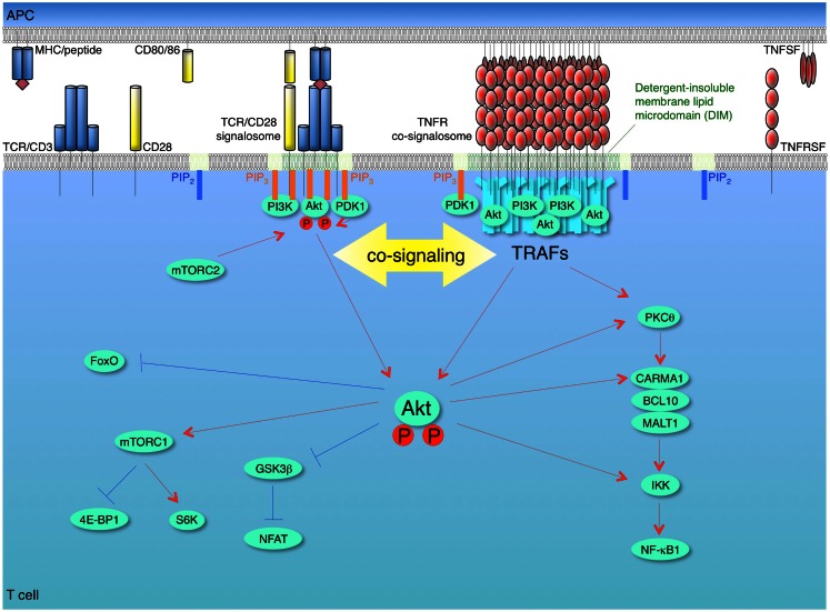
Figure 2.
Model of synergy between TCR/CD28 and TNFR signalosomes for activation of the PI3K-Akt pathway in T cells. T cells are activated firstly by recognition of antigen by the T cell receptor (TCR)/CD3 complex when it is displayed by the major histocompatibility complex (MHC) on antigen-presenting cells (APCs). The second co-stimulatory signal is delivered through CD28 by interaction with its ligands CD80 and/or CD86. These combined signals can activate phosphoinositide 3-kinase (PI3K), which leads to conversion of PIP2 into PIP3 at the plasma membrane. The pleckstrin homology (PH) domain containing proteins, Akt (protein kinase B) and phosphoinositide-dependent kinase 1 (PDK1), are recruited to the membrane PIP3, and then Akt is phosphorylated by PDK1 and by the mammalian target of rapamycin complex 2 (mTORC2). This promotes translocation of Akt from membrane to cytosol, thereby allowing regulation of downstream pathways through phosphorylation of target molecules, such as glycogen synthase kinase 3β (GSK3β), forkhead box O (Foxo), and IκB kinase (IKK). Akt phosphorylates and inactivates two negative regulators of mTORC1, tuberous sclerosis complex 2 (TSC2) and proline-rich Akt substrate of 40 kDa (PRAS40), which results in activation of mTORC1. Akt contributes to NF-κB activation through phosphorylation of IKK and interaction with protein kinase C θ (PKCθ) or caspase-recruitment domain (CARD)-membrane-associated guanylate kinase (MAGUK) protein 1 (CARMA1). After recognition of trimeric TNF ligand superfamily (TNFSF) molecules on APCs, TNF receptor superfamily (TNFRSF) molecules on T cells are trimerized and oligomerized and recruit trimeric TNFR-associated factors (TRAFs) to their cytoplasmic TRAF-binding motifs. The TNFSF-TNFRSF complex then translocates into detergent-insoluble membrane lipid microdomains (DIM). The TNFRSF-TRAF superclusters recruit and allow the efficient accumulation of PI3K and Akt in concentrated depots in close proximity to the TCR/CD28 signalosome, which results in a quantitative contribution of TNFR signalosomes to enhancing and sustaining PI3K and Akt activation triggered by the TCR/CD28 signalosome. The TNFR signalosomes also can promote activation of NF-κB irrespective of TCR/CD28 signaling. Red lines show activating signals, blue lines show inhibitory signals.

