Figure 2.
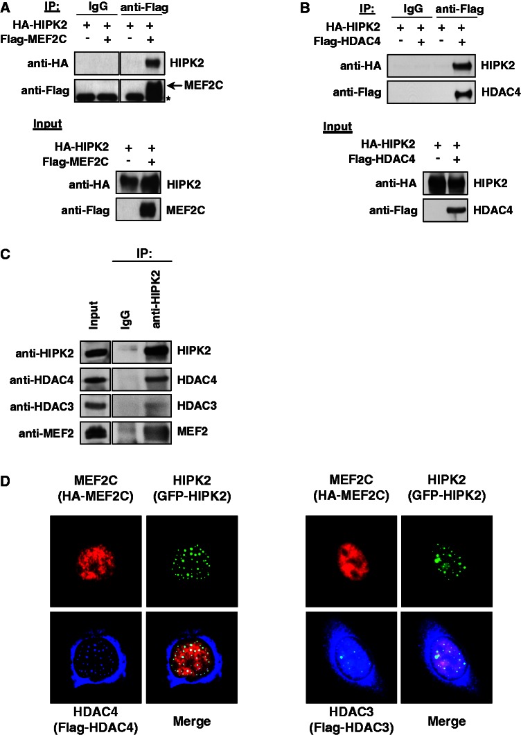
HIPK2 interacts with MEF2C and HDAC4. (A) Epitope-tagged versions of MEF2C and HIPK2 were coexpressed in 293T cells. A fraction of the cell lysates was tested for the correct expression of the transfected proteins by immunoblotting (lower), while the remaining extracts were used for IP with anti-Flag antibodies or isotype-matched controls. After elution of bound proteins in 1× SDS sample buffer, coprecipitated HIPK2 was visualized by western blotting as shown. An asterisk indicates a non-specific band. (B) The experiment was done as in (A) with the exception that Flag-HDAC4 was expressed instead of Flag-MEF2C. (C) Lysates from C2C12 cells were used for IP with anti-HIPK2 antibodies or adequate controls. The coprecipitating endogenous MEF2 proteins, as well as HDAC3 and HDAC4, were revealed by immunoblotting as shown. (D) U2OS cells were transfected to express the indicated proteins and further analyzed by indirect immunofluorescence for the intracellular distribution of HIPK2 and its interaction partners. The merged pictures display colocalizing proteins in the white areas; representative pictures are displayed.

