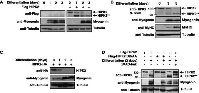
Figure 5.
HIPK2 is cleaved by caspases during myogenic differentiation. (A) C2C12 cells were infected with lentiviruses to express Flag-HIPK2 as shown, followed by the induction of differentiation for the indicated periods. Protein extracts were analyzed by western blotting for the expression of myogenin and HIPK2 with specific antibodies. The shorter HIPK2 form is indicated as HIPK2**. (B) C2C12 myoblasts were stimulated with differentiation medium to enter the differentiation program and harvested at different time points. Cell extracts were analyzed by immunoblotting to reveal the differentiation state, as revealed by increased expression of myogenin and MyHC. HIPK2 processing was analyzed with N6A10 antibodies and the full-length and cleaved forms are shown. The positions of molecular weight marker proteins are displayed. (C) C2C12 cells were transfected to express HIPK2 fused to a C-terminal HA-tag, followed by the induction of differentiation and western blot analysis as shown. (D) C2C12 cells were transfected to express Flag-HIPK2 or Flag-HIPK2 DD/AA as shown. Muscle differentiation was induced for 2 days in the absence or presence of the caspase inhibitor zVAD-fmk (20 μM added for the last 12 h before harvest). Cell lysates were tested for HIPK2 cleavage and expression of myogenin with specific antibodies.

