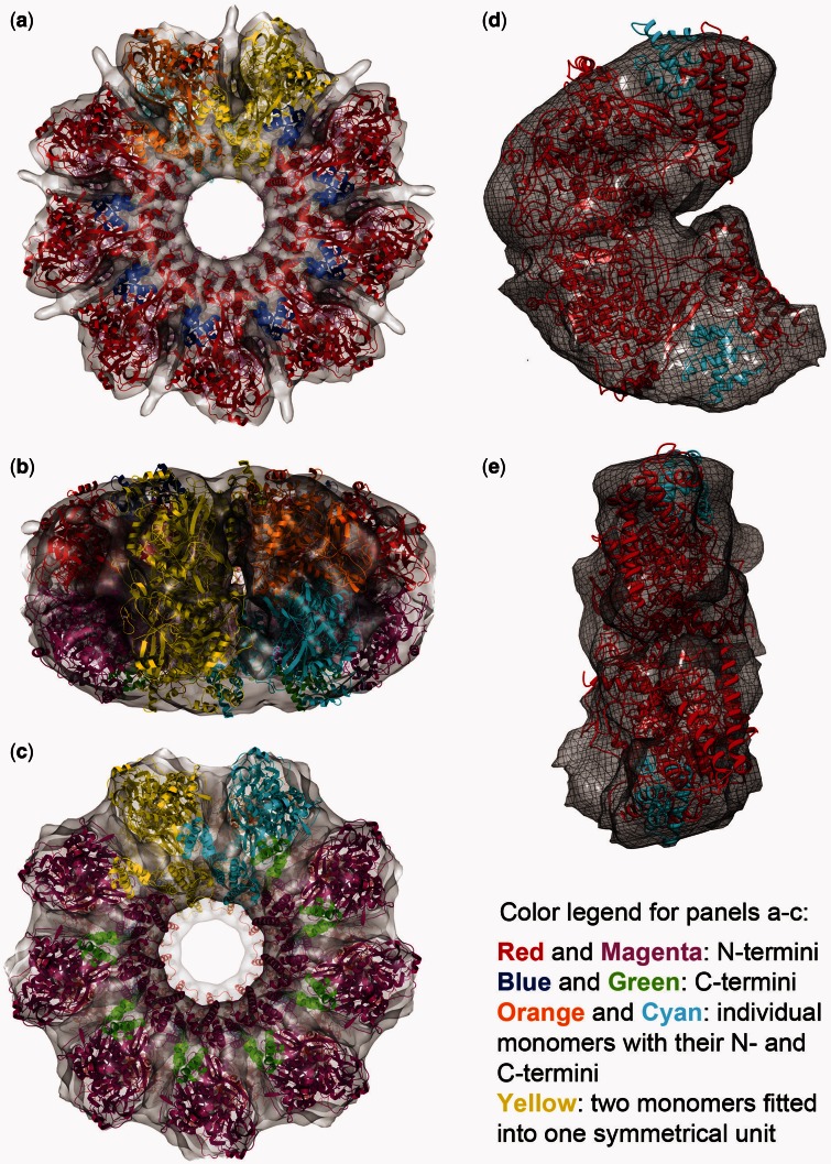
Figure 3.
Fitting the X-ray crystal structure. Eighteen copies of a C-terminal truncated ICP8 monomer [(28) and ‘Materials and Methods’ section] were fit into the reconstructed double-ring map, nine on top and nine at the bottom. (a–c) They are the top, side and the bottom views, respectively; red and magenta are the N-terminal domains, and blue and green are the C-terminal domains. Cyan and orange monomers were colored such that to show individual monomers with their N- and C-termini. Yellow shows two monomers fitted into one symmetrical unit. (d and e) They show one symmetrical unit containing two monomers isolated from the map. d shows the side view facing the next symmetrical unit, and e shows the side that faces to the center of the ring. Red and cyan are the N- and C-terminal domains, respectively, fitted into one symmetrical unit composed of top and bottom densities.

