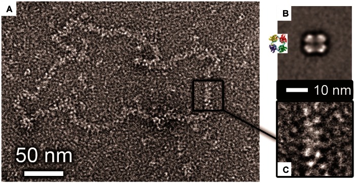
Figure 4.
EM of purified tetramers and native RNPs. (A) Micrograph of negative-stained RNP purified from BUNV virus particles in cell culture. (B) Single particle average of purified SBV tetramers bound to synthetic 48mer RNA (inset shows the crystal structure of the BUNV N tetramer bound to RNA). (C) Zoomed in view of a section of native RNP at the same scale as B (the 10-nm scale bar applies to both B and C).

