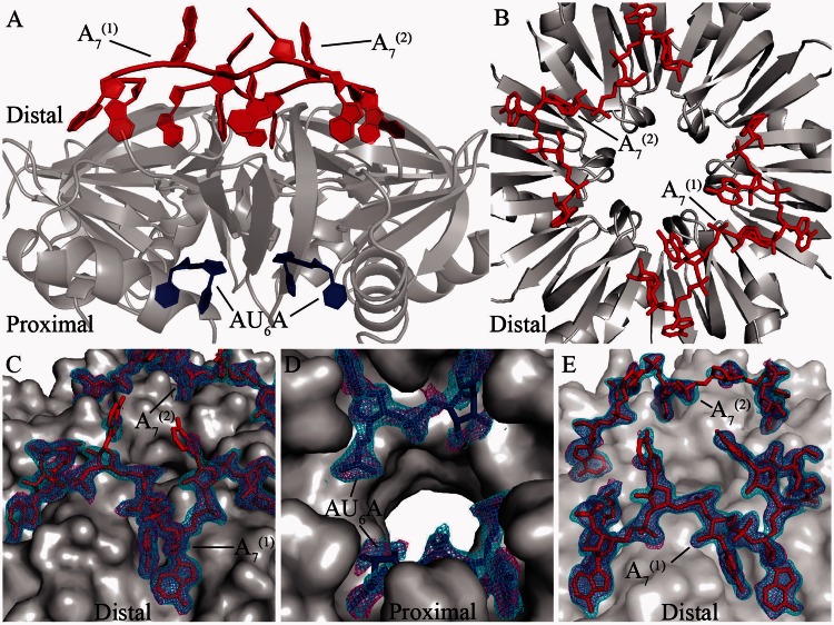
Figure 2.
Global structures of AU6A•Hfq•A7 and Hfq•A7 complexes. In the AU6A•Hfq•A7 crystal, each asymmetric unit contained half of the Hfq hexamer. Biologically relevant assembly was generated according to crystallographic symmetry. (A) In the AU6A•Hfq•A7 structure, two A7 (red) molecules and one AU6A (blue) molecule are bound to each Hfq hexamer (gray) on distal and proximal sides, respectively. (B) Two A7 (red) molecules are bound to distal side of Hfq hexamer (gray) in the Hfq•A7 structure. (C) Clear density maps are observed for the two A7 molecules (red) in the AU6A•Hfq•A7 structure. (D) Part of AU6A in the AU6A•Hfq•A7 structure. (E) Electron densities for the two A7 molecules in the Hfq•A7 structure are also clearly observed. Difference maps Fo–Fc before inclusion of RNAs are shown as purple mesh (contoured at 2.0 σ), and 2Fo–Fc densities are shown as cyan mesh (contoured at 1.0 σ). The statistics of these two structures are shown in Supplementary Table S1.

