Figure 2.
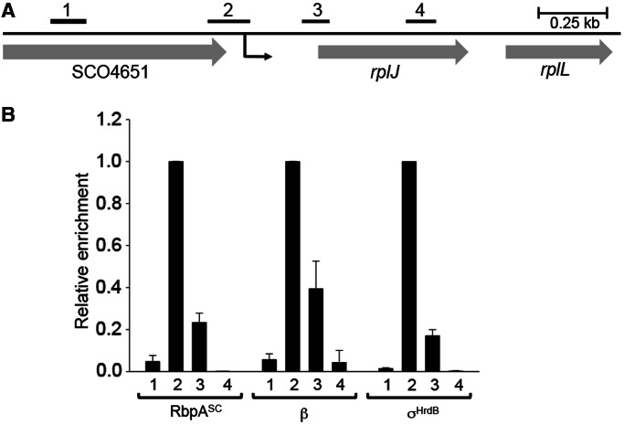
Localization of RbpASc at a σHrdB-dependent promoter in vivo. (A) Schematic of the rplJ region. The transcription start point (+1) of rplJp is indicated by a black arrow located 240 bp upstream from the rplJ start codon. The bars above the genes show the relative positions of PCR products used for ChIP–qPCR that are centred with respect to +1 as follows: 1, −653 bp; 2, −79 bp; 3, +235 bp; 4, +235 bp. (B) Occupancy of RbpASc, RNAP β subunit, and σHrdB at the indicated regions in S. coelicolor S129 (pSX190), after treatment with rifampicin. Immunoprecipitations were performed using monoclonal anti-β, polyclonal anti-σHrdB and anti-FLAG antibody to detect RbpA–FLAG. To allow comparison of RbpASc, β and σHrdB localization, after absolute quantitation of co-immunoprecipitated DNA and background correction for each antibody, enrichment is presented relative to the highest corrected signal obtained using either of the four primer pairs. Standard deviations (calculated for two biological replicates) are indicated.

