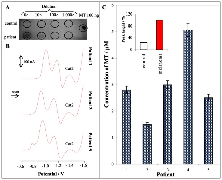
Figure 4.
Immunological detection of MT on PVDF membrane, sample volume: 1 μl, visualization: AEC (A). Typical DP voltammograms of blood serum from patients with malignant melanoma (patient 1, 3 and 5) (B). MT content in patients with malignant melanoma, in inset: MT level at healthy persons (n = 20) and patients with malignant melanoma (n = 5) (C). The samples were processed according to procedure mentioned in “Materials and Methods” section. The measurement of 5 μl of 1000 times diluted sample by 0.2 M phosphate buffer (pH 6.8) was performed by AdTS DPV Brdicka reaction.

