FIGURE 7.
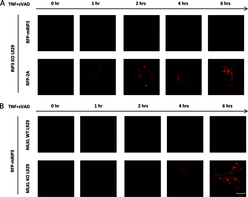
Loss of interaction with MLKL leads to massive aggregation of RIP3 in TNF-stimulated L929 cells. A, RIP3 KO L929 cells were infected with lentivirus encoding RFP-mRIP3 or RFP-mRIP3–2A. The infected cells were then treated with TNF plus Z-VAD for different periods of time as indicated and live cell imaging was recorded. B, the same as in A except that MLKL wild-type and KO L929 cells were infected with lentivirus encoding RFP-mRIP3. Scale bar, 10 μm.

