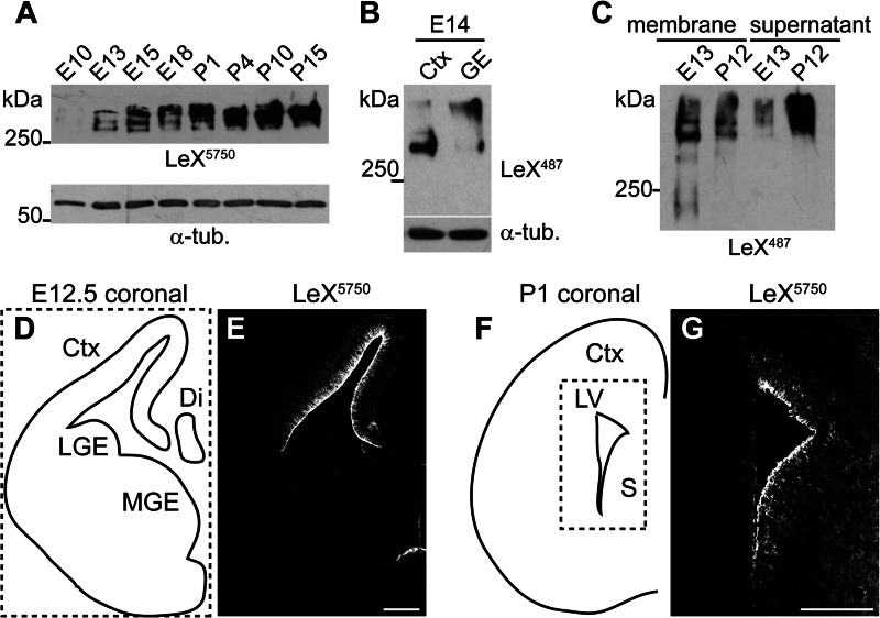
FIGURE 1.
LeX carrier proteins change during mouse CNS development. A, Western blot with anti-LeX mAb 5750LeX of brain lysates at the indicated developmental stages. Note that the detected LeX-positive proteins shift from embryonic to postnatal stages. B, Western blot against LeX of E14 cortex (Ctx) and ganglionic eminence (GE). C, at E13 most LeX-positive proteins are membrane-associated, whereas at P12 LeX proteins accumulate in the membrane-free (supernatant) fraction after differential centrifugation. D–G, LeX immunostainings of E12.5 (E) or P1 (G) coronal forebrain sections depicted in D and F. L/MGE, lateral/medial GE; Di, diencephalon; LV, lateral ventricle; S, septum, α-tub., α-tubulin. Scale bars, 200 μm

