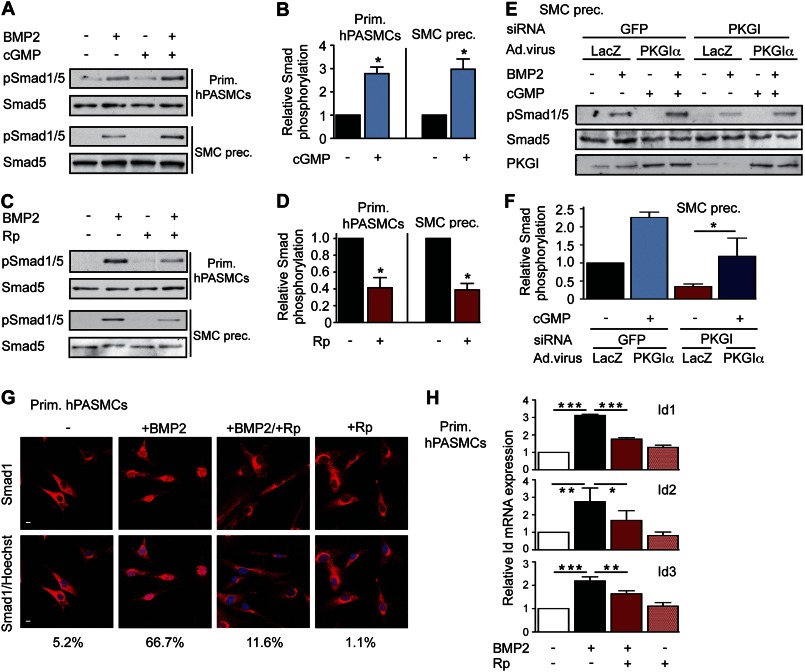
FIGURE 1.
PKGI enhances Smad phosphorylation and downstream signaling in SMCs. A and B, primary (Prim.) human PASMCs and SMC precursors (SMC prec.) were treated with 3 nm BMP2 and/or 100 μm 8-pCPT-cGMP (cGMP) for 30 min. Smad1/5 phosphorylation was examined by immunoblotting, and three experiments were quantified, with BMP2-induced Smad phosphorylation assigned a value of 1; *, p < 0.05, two-tailed Student's t test. Error bars, S.E. C and D, cells were treated with BMP2 and/or 50 μm Rp-pCPT-PET-cGMPS (Rp), and Smad activation was analyzed as in A and B (*, p < 0.05). E and F, SMC precursors were transfected with siRNAs targeting GFP (control) or PKGI, and PKG was reconstituted by adenoviral vector encoding siRNA-resistant PKGIα. Cells were treated and analyzed as in A and B, with BMP2-induced Smad phosphorylation of GFP siRNA/LacZ virus-treated cells assigned a value of 1 (*, p < 0.05, one-way ANOVA). PKGI expression was assessed by immunoblotting. G, human PASMCs were treated as in C, and Smad1 nuclear translocation was examined by immunofluorescence labeling (red); nuclei were counterstained with Hoechst (blue). Scale bars, 5 μm. H, human PASMCs were treated with BMP2 and/or Rp for 4 h, and Id1/2/3 mRNA levels were quantified by real-time RT-PCR. After normalization to Gapdh, relative Id mRNA in untreated cells was assigned a value of 1 (*, p < 0.05; **, p < 0.01; ***, p < 0.001, one-way ANOVA).

