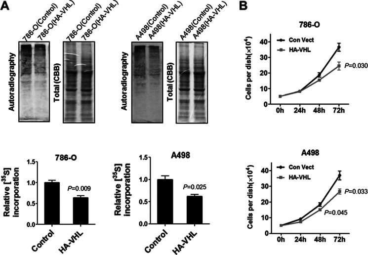
FIGURE 6.
pVHL inhibits protein synthesis. A, pVHL expression inhibits protein synthesis. 786-O and A498 cells that were stably transfected with HA-VHL were seeded in 12-well plates. The next day, the medium was removed and methionine-free medium was added. After 45 min, the cells were provided with [35S]methionine (10 μCi/ml) and incubated for 60 min. The cells were harvested and lysed. Cellular proteins were resolved on SDS-PAGE and the 35S-labeled proteins were visualized by autoradiography. 786-O and A498 cells stably transfected with control vector were used as control. The 35S-incorporation was determined by measuring the density of the 35S-labeled protein (autoradiograph) and normalized to that of total proteins (Coomassie Blue stain). The data are mean ± S.E. (n = 3). B, re-expression of pVHL inhibits cell proliferation. 786-O and A498 cells stably expressing pVHL were selected as described under “Materials and Methods.” Cell proliferation was determined by counting the cell numbers using a hemocytometer. The data are mean ± S.E. (n = 3).

