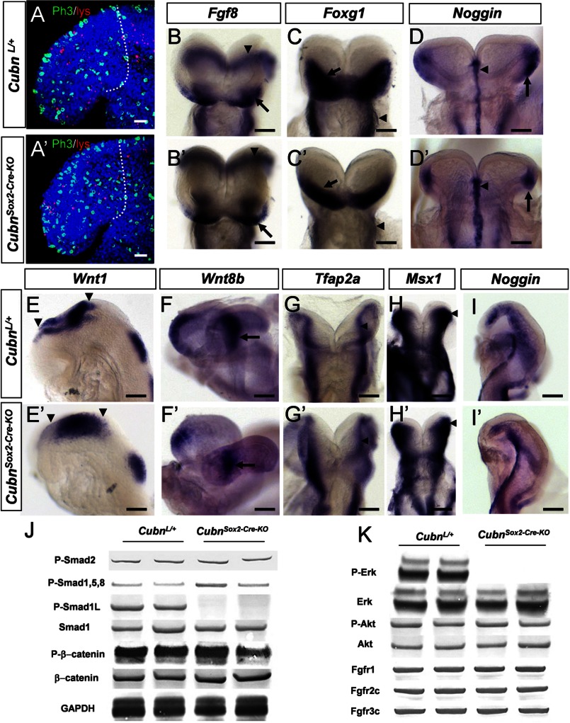
FIGURE 4.
Normal forebrain regionalization and impaired MAPK signaling in E8.75 CubnSox2-Cre-KO mutant heads. A and A′, representative lateral views of E8.75 control and CubnSox2-Cre-KO embryos stained with Lysotracker (in red) and anti-phospho-histone H3 (in green). Dotted lines demarcate the forebrain region in confocal sections of an 8-ss control (A) and mutant (A′) embryo. Nuclei colored by Hoechst are shown in blue. B–I, representative whole-mount mRNA staining of control (B–I) and mutant (B′–I′) embryos at E8.75. B and B′, normal Fg8 expression in the ANR (arrow) and the isthmus (arrowhead). C and C′, Foxg1 is expressed in the anterior neural plate (arrow); it is reduced in the mutant pharyngeal region (arrowhead). D and D′, Noggin is normally detected in the rostral axial mesendoderm (arrowhead) and the rostrolateral ectoderm (arrow). E–F′, normal expression of Wnt1 in the developing midbrain (arrowheads in E and E′) and of Wnt8b in the developing caudal forebrain (arrow in F and F′). G–H′, dorsal views of control and mutant embryos at the level of the prospective midbrain (arrowhead) and hindbrain showing Tfap2a normally expressed in the dorsal neural folds (G and G′) and Msx1 in the dorsal neural folds and cephalic mesenchyme (H and H′). I and I′, Noggin is similarly found in a lateral strip of cells between the rostral-most cephalic mesenchyme and the border of the rhombencephalon (r2–r3). Frontal views (B–D′); lateral views with the anterior to the left (E–F′ and I and I′). J, immunoblot analysis of cephalic extracts for phospho-Smad2, phospho-Smad1/5/8, phospho-Smad1Linker, total Smad1, phospho-β-catenin, and β-catenin. GAPDH is used as loading control. K, phospho-ERK1/2 is decreased, and phospho-Akt is maintained. The levels of endogenous total ERK and Akt are shown for comparison. The levels of FgfR1, FgfR2c, and Fgfr3c are normal in the mutants. Scale bars, A, 30 μm; B–D, 100 μm; E, H, and I, 6 μm; F and G, 40 μm.

