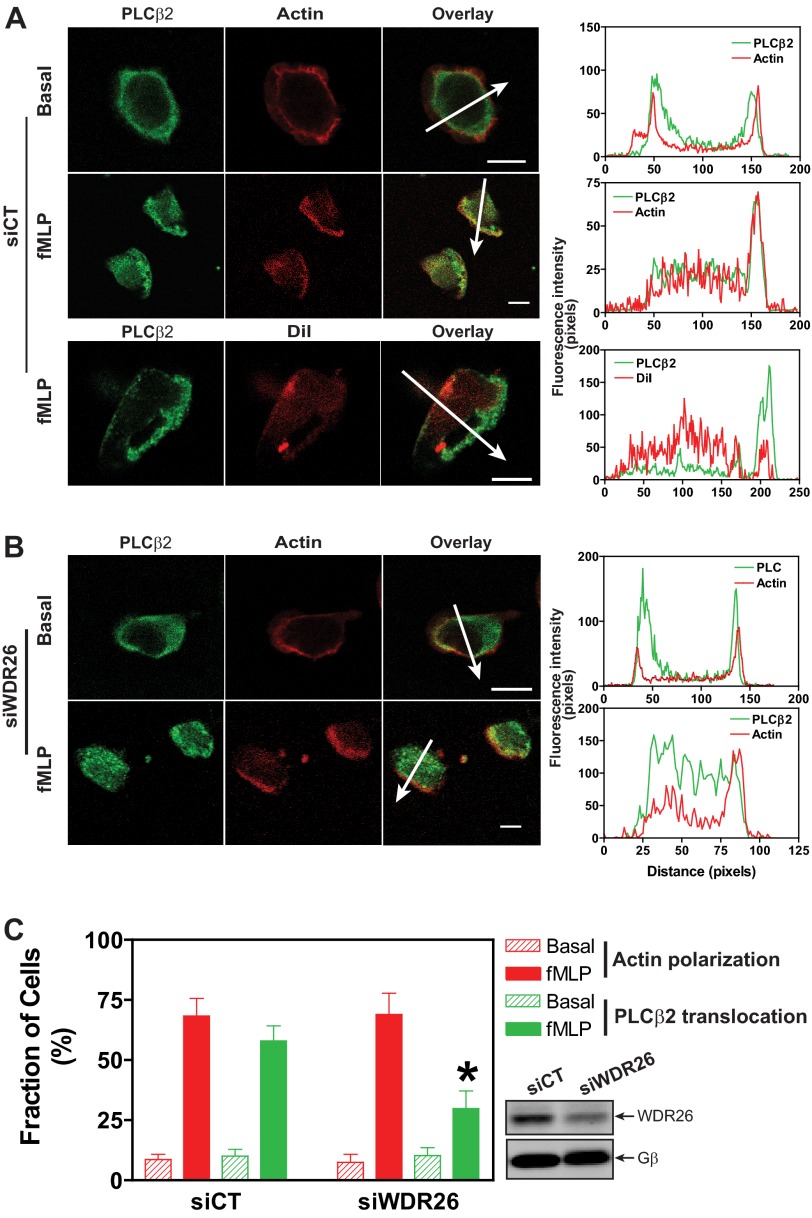
FIGURE 6.
WDR26 regulates PLCβ2 translocation. dHL60 cells were transiently transfected with a control (siCT) (A) or WDR26 siRNA (siWDR26) (B) and stimulated with buffer (Basal) or fMLP (0.2 μm) for 2 min. After fixation and permeabilization, cells were stained with a rabbit anti-PLCβ2 antibody and Alexa Fluor 568-conjugated phalloidin or CM-DiI. Representative images are shown in A and B, and quantitative data from over 100 cells in three separate experiments are shown in C. *, p < 0.05 versus siCT. The graphs in the right panel show the distribution of fluorescence intensity of PLCβ2 and F-actin or CM-DiI along the arrows drawn across the cells. Bar, 10 μm. The level of WDR26 and Gβ expression in control and WDR26 siRNA cells is shown in representative blots in C. Error bars represent S.E.

