
FIGURE 4.
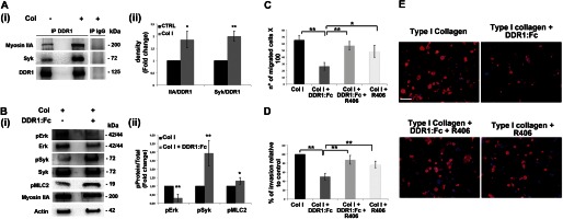
Syk kinase is involved in DDR1-mediated MK migration on type I collagen. A, panel i, DDR1 was immunoprecipitated (IP) in cell lysates of MKs plated for 16 h on type I collagen (Col) or on tissue culture plastic. A control sample was immunoprecipitated with an unrelated antibody (IgG). Membranes were probed with anti-Syk and anti-myosin IIA antibodies to show DDR1-interacting protein and reprobed with anti-DDR1 antibody to show equal loading. Panel ii, densitometry analysis of the Western blots of Myosin IIA and Syk co-immunoprecipitated with DDR1. B, total cellular lysates of MKs plated for 16 h on type I collagen, inhibiting or not inhibiting DDR1 activation, were subjected to Western blot analysis. Membranes were probed with the indicated antibody, with p indicating the phosphorylated form. Actin was probed to show equal loading. Panel ii, densitometry analysis of the Western blots of phospho-ERK (pERK), phospho-Syk (pSyk), and phospho-MLC2 (pMLC2). C, Transwell migration assay of mature MKs through type I collagen, in the presence of the DDR1-Fc blocking molecule and of Syk specific inhibitor compound R406 (5 μm) either mixed or used singularly. After 16 h, MKs that had passed in the lower chamber were counted by phase contrast microscopy. D, MKs adhering to the lower side of the Transwell filter were fixed and stained with anti-CD61 antibody and then counted by fluorescence microscopy. E, representative images of MK invasion of type I collagen. Cells adhering to the lower side of the Transwell coated filter were fixed and stained with anti-CD61 antibody (red). (Immunofluorescence staining, Olympus BX51 microscope, magnifications 20×.) Scale bars are 100 μm. Nuclei were stained with Hoechst 33288 (blue). Data are presented as means ± S.D.(n = 5, 5, and 5 independent experiment). *, p < 0.05. **, p < 0.01.