
FIGURE 5.
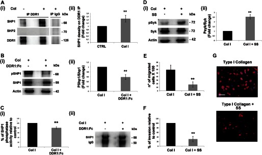
SHP1 modulates Syk activation upon type I collagen-DDR1 binding in MKs. A, panel i, DDR1 immunoprecipitates (IP) from MKs plated for 16 h on type I collagen (Col) or on plastic were analyzed by Western blot. A control sample was immunoprecipitated with an unrelated antibody (IgG). Membranes were probed with anti-SHP1 and anti-SHP2 antibodies to show the co-immunoprecipitation and with anti-DDR1 antibody to show equal loading. Panel ii densitometry analysis of the Western blots of SHP1 protein co-immunoprecipitated with DDR1. B, panel i, Western blot analysis of cell lysates of MKs plated for 16 h on type I collagen mixed with or without DDR1-Fc blocking molecule. Membranes were probed with the indicated antibodies with p indicating the phosphorylated form. Actin was probed to show equal loading. Panel ii, densitometry analysis of the Western blots of pSHP1. CTRL, control. C, panel i, SHP1 phosphatase activity measured in a phosphatase assay on SHP1 immunoprecipitates from MK lysates, using p-nitrophenyl phosphate as substrate. MKs were plated for 16 h on type I collagen mixed with or without DDR1-Fc blocking molecule. Panel ii, Western blot analysis of SHP1 immunoprecipitates used for SHP1 phosphatase activity assay. SHP1 phosphatase activity was related to the same concentration of SHP1. Shown here is a representative Western blot out of four independent experiments. D, panel i, MKs were treated with the SHP1 specific inhibitor sodium stibogluconate (SS) (13.4 μm) and plated for 3 h on type I collagen. Cell lysates were subjected to Western blot analysis. Membranes were probed with anti-phospho Syk (Tyr-525/526) antibody and anti-Syk and anti-actin antibodies to show equal loading. Panel ii, densitometry analysis of the Western blots of phospho-Syk (pSyk). E, Transwell migration assay of mature MKs through type I collagen, in the presence of the SHP1 specific inhibitor SS (13.4 μm). After 16 h, MKs that had passed in the lower chamber were counted by phase contrast microscopy. F, MKs adhering to the lower side of the Transwell filter were fixed and stained with anti-CD61 antibody and then counted by fluorescence microscopy. G, representative images of MK invasion of type I collagen. Cells adhering to the lower side of the Transwell coated filter were fixed and stained with anti-CD61 antibody (red) antibody. (Immunofluorescence staining, Olympus BX51 microscope, magnifications 20×.) Scale bars are 100 μm. Nuclei were stained with Hoechst 33288 (blue). Data are presented as means ± S.D. (n = 5, 4, 4, 4, 3, and 3 independent experiments). *, p < 0.05 **, p < 0.01.