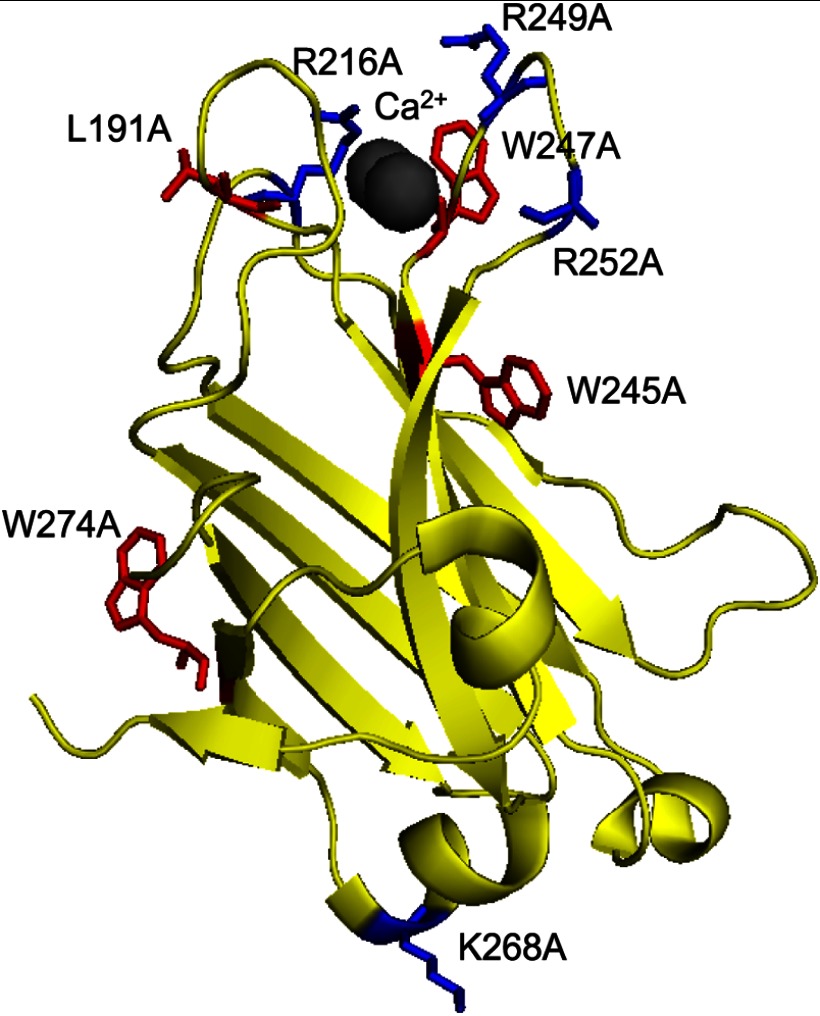
FIGURE 1.
Structure of the C2 domain of PKCα. A ribbon diagram of the C2 domain of PKCα (based on the structure by Gómez-Fernández and colleagues (22)) shows residues mutated in this study. Basic residues are indicated in blue (Arg-216, Arg-249, and Arg-252 on the Ca2+-binding loops and the distally located Lys-268), and hydrophobic residues are indicated in red (Leu-191, Trp-247, and Trp-245 on the Ca2+-binding loops and the distally located Trp-274).

