
FIGURE 7.
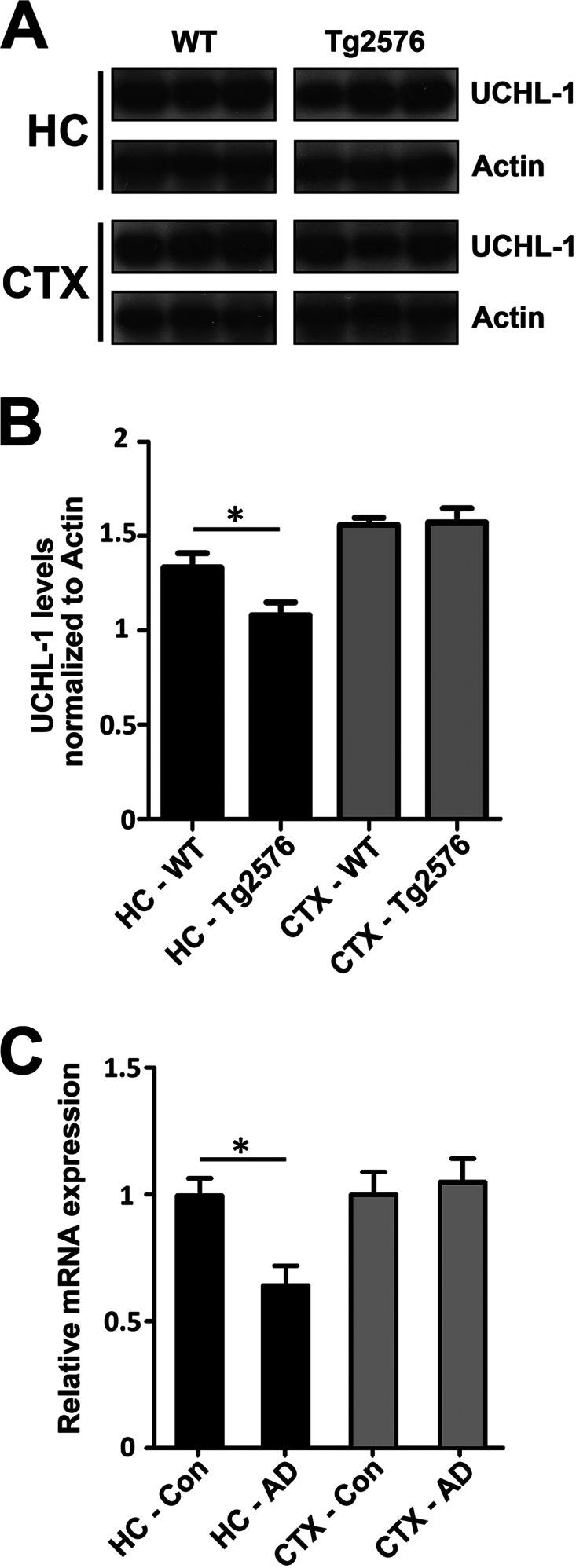
UCH-L1 is decreased in Tg2576 mice and in the Alzheimer disease brain. A, levels of UCH-L1 are decreased in APP-Tg2576 hippocampus. Hippocampal lysates were prepared as described under “Experimental Procedures.” Protein was separated on SDS-PAGE, and Western blot analysis was carried out to determine the amount of UCH-L1 protein in wild-type and Tg2576 mouse brain. B, UCH-L1 protein levels are decreased within the hippocampus but not the cortex of 15-month-old Tg2576 mice (*, p < 0.03). UCH-L1 levels were quantitated and normalized to actin protein as described under “Experimental Procedures.” C, UCHL-1 gene expression is lower in AD brain. Expression profiles were obtained from a microarray database consisting of brain tissue from AD cases (n = 26; range, 74–95 years; mean age, 85.7 ± 6.5 years) and age-matched controls (n = 33; range, 69–99 years; mean age, 84.2 ± 8.9 years) and were generated using Affymetrix HgU133 plus 2.0 arrays as described previously (44). Two probe sets corresponding to UCHL-1 (Unigene Hs.518731) were identified on the HgU133 plus 2.0 array, both of which had Present flags in all microarrays, indicating high expression reliability of the probes. Expression values were averaged across the probe sets to obtain an overall value for each case, followed by t test comparisons for each region and significance set at p < 0.05 (*).