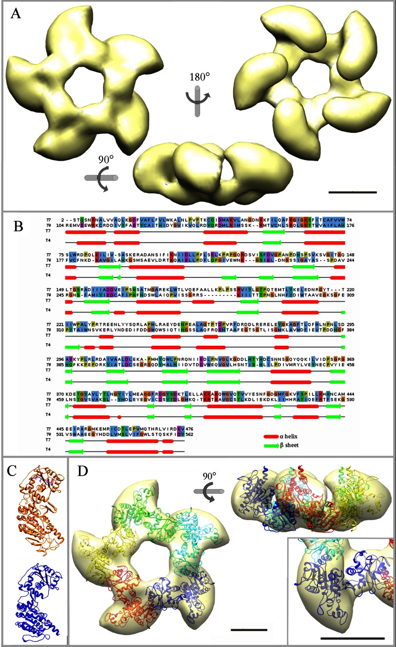
FIGURE 2.
Three-dimensional reconstruction of the pentameric large terminase and fitting of the gp19 atomic model. A, shown is an EM reconstruction of the pentameric terminase. The structure is shown in three different orientations, as indicated. B, sequence alignment used for the modeling of the T7 large terminase (gp19, target protein) based on the template structure of the T4 large terminase (gp17, 3CPE) shows conserved motifs at the amino acid level and the secondary structures correspondence. C, shown is a comparison of the gp17 atomic structure (in orange) and the gp19 final model (in blue). D, shown is a translucent model of the terminase structure together with the fitted atomic model of the gp19 pentamer in end-on and side views. Each gp19 monomer is presented in a different color. Inset, two detailed views of the fitted monomer in different orientations. The scale bar represents 50 Å.

