FIGURE 3.
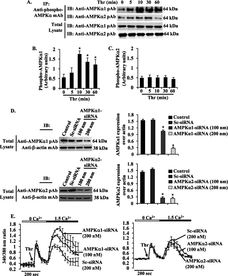
PAR-1-activated AMPKα1 regulates SOCE in HLMVECs. A, HLMVECs were challenged with thrombin (50 nm) for different time intervals at 37 °C. After thrombin stimulation, cells were lysed and immunoprecipitated (IP) with anti-phospho-AMPKα mAb. The precipitated proteins were immunoblotted (IB) with anti-AMPKα1 pAb (1st lane) or AMPKα2 pAb (2nd lane). Total cell lysates were immunoblotted with anti-AMPKα1 pAb (3rd lane) or AMPKα2 pAb (4th lane). Results are shown as the mean ± S.E. of four independent experiments for AMPKα1 (B) or AMPKα2 (C). * indicates the significance compared with cells not treated with thrombin. D, HLMVECs transfected with Sc-siRNA, AMPKα1-siRNA, or AMPKα1-siRNA (see details under “Experimental Procedures”) were lysed and immunoblotted with anti-AMPKα1 pAb (left, top panel) or anti-AMPKα2 pAb (left, bottom panel). The membrane was stripped and probed with anti-β-actin mAb as loading control. In right panels, AMPKα1 and AMPKα2 proteins were quantified by densitometry relative to β-actin. Results shown are mean ± S.E. of four experiments. *, significantly different compared with control or Sc-siRNA transfected cells. E, HLMVECs transfected with Sc-siRNA, AMPKα1-siRNA (100 or 200 nm), or AMPKα2-siRNA (100 or 200 nm) were used to measure thrombin (Thr)-induced Ca2+ store release and Ca2+ entry as described in Fig. 2C. Arrow indicates the time when the cells are challenged with thrombin (Thr). Results shown are mean ± S.E. of four experiments.

