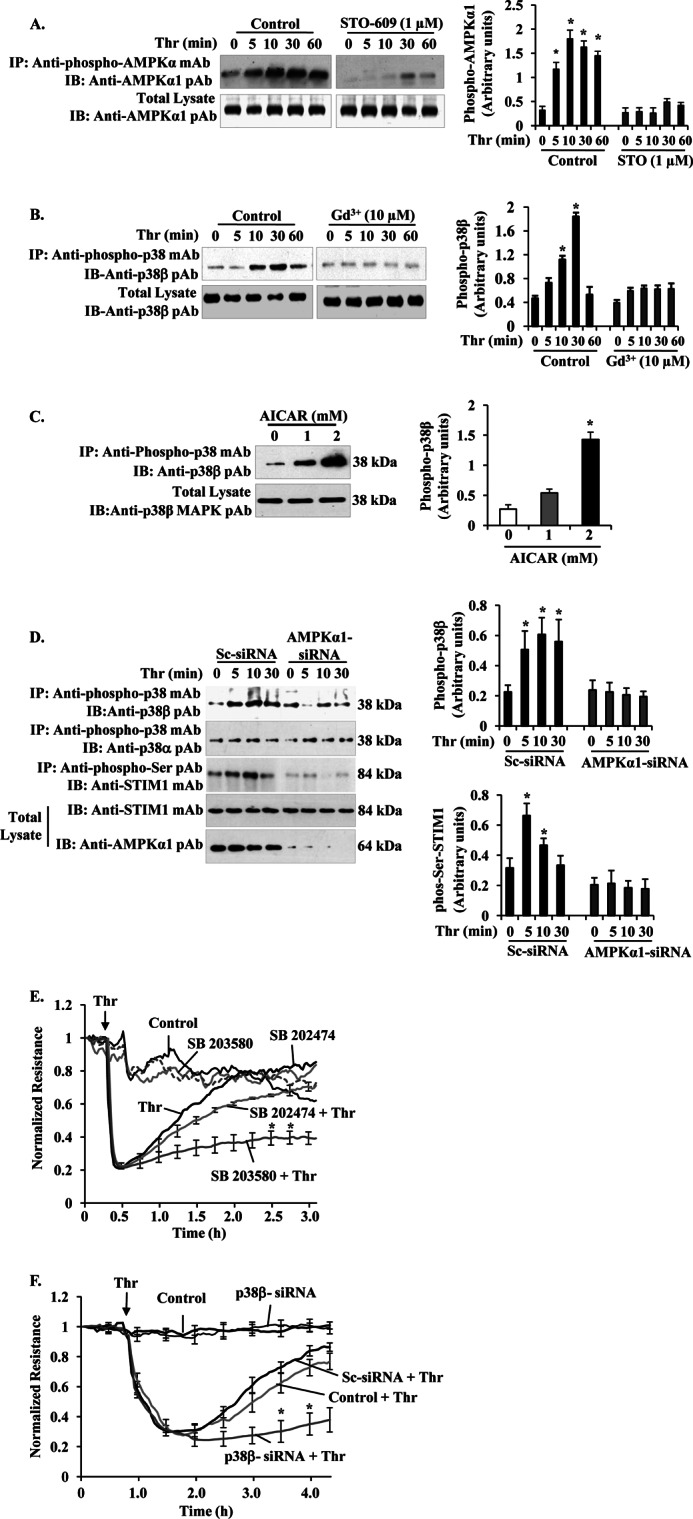
FIGURE 6.

Ca2+ entry-CaMKKβ-AMPKα1-p38β MAPK axis signaling mediates STIM1 phosphorylation to inhibit SOCE and endothelial permeability. A, HLMVECs were pretreated with or without CaMKKβ inhibitor STO-609 (1 μm) for 30 min, and then thrombin-induced AMPKα1 phosphorylation was measured as described in Fig. 3A. The experiment was repeated four times, and the results shown are mean ± S.E. (right panel). *, significantly different from cells not stimulated with thrombin. B, HLMVECs, thrombin-induced p38β phosphorylation was measured in the absence and presence of Gd3+ (10 μm). After thrombin treatment, cell lysates were immunoprecipitated (IP) with anti-phospho-p38 mAb, and the precipitate was immunoblotted (IB) with anti-p38β pAb to determine p38β phosphorylation (top panel). Total cell lysates were blotted with anti-p38β mAb (bottom panel). A representative blot is shown from four independent experiments. Phosphoprotein bands were quantified by densitometry and are expressed in arbitrary units. *, significantly different from cells not stimulated with thrombin. Note that impaired thrombin-induced p38β phosphorylation in cells treated with Gd3+ to inhibit Ca2+ entry. C, HLMVECs pretreated with AICAR (0, 1, and 2 mm) were lysed and immunoprecipitated with anti-phospho-p38 mAb. The precipitated proteins were immunoblotted with anti-p38β MAPK pAb. Total cell lysates were immunoblotted with anti-p38β MAPK pAb. D, HLMVECs transfected with either Sc-siRNA or AMPKα1siRNA (200 nm) as described in Fig. 3D were stimulated with thrombin (50 nm) for different time intervals at 37 °C. After thrombin treatment, cells were lysed and immunoprecipitated (IP) with anti-phospho-p38 mAb. The precipitated proteins were immunoblotted (IB) with anti-p38β pAb (top row) or anti-p38α pAb (2nd row). Total cell lysates were immunoprecipitated with anti-phospho-Ser pAb, and the precipitate was immunoblotted with STIM1 mAb (3rd row). Total cell lysates were immunoblotted with anti-STIM1 mAb (4th row) and anti-AMPKα1 pAb (bottom row). Results shown are the mean ± S.E. of four independent experiments (right panels). *, significantly different from control cells. E, HLMVECs were grown to confluence on gold electrodes (see details under “Experimental Procedures”). Cells were washed and incubated with 1% serum-containing medium for 2 h and then incubated 30 min with or without the indicated concentrations of 10 μm SB203580 or SB202474 before the addition of 50 nm thrombin (Thr). Note that in SB203580-treated cells, thrombin produced a marked decrease in TER, but the TER recovery to basal level was delayed compared with control cells treated with thrombin or cells pretreated with SB202474 followed by thrombin addition. The arrow indicates the time at which the cells were challenged with thrombin (Thr) or medium. F, HLMVECs transfected with 200 nm Sc-siRNA or p38 β-siRNA were used to measure thrombin-induced TER changes. Note the delay in thrombin-induced decrease in TER recovery to basal level in p38β-siRNA transfected cells indicating hyper-permeability associated with prolonged SOCE. The arrow indicates the time at which the cells were challenged with thrombin (Thr) or medium. Results shown are the mean ± S.E. of four independent experiments. *, significantly different from control cells treated with thrombin.
