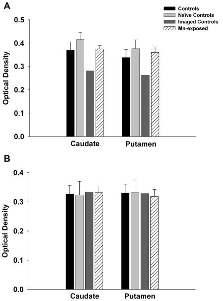
Figure 6.
Tyrosine hydroxylase (TH) and dopamine transporter (DAT) Immunohistochemistry. Panel A shows optical density readings for DAT in the caudate and putamen. Consistent with the autoradiography data, DAT levels were reduced in the imaged-controls that received AMPH only as part of the imaging protocol. However, DAT levels were not affected by chronic Mn exposure. Similarly, neither Mn-exposure nor PET-related AMPH treatment affected TH levels (Panel B). Each value is the mean ± sem of n=6 (controls), n=4 (naïve controls), n=2 (imaged controls), and n=7 (Mn-exposed).

