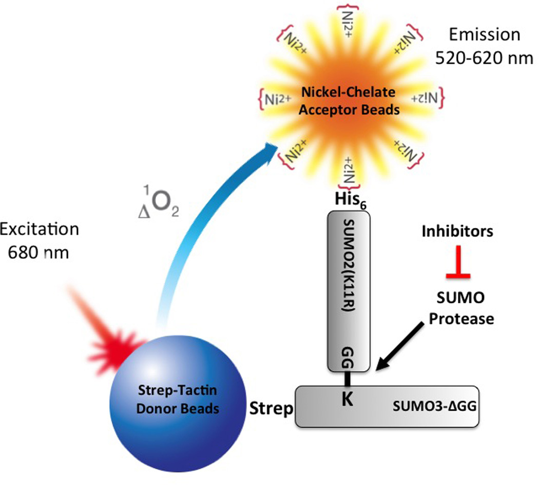
Figure 1.

Principle of the AlphaScreen-based SUMO protease inhibitor assay. In this assay format, His-SUMO2(K11R) conjugated to Strep-SUMO3ΔGG (SS3HS2) is used as the SUMO protease substrate. When substrate SS3HS2 is mixed with Strep-Tactin donor beads and Nickel Chelate acceptor beads, donor beads and acceptor beads bind to Strep-SUMO3ΔGG and His-SUMO2(K11R), respectively. Upon laser excitation, donor beads generate singlet oxygen that diffuses to and activates acceptor beads when in close proximity, resulting in signal emission. When the substrate is cleaved by SUMO protease, the distance between His tag and Strep tag is increased, resulting in a marked decrease in signal intensity due to a short lifetime of singlet oxygen in aqueous solutions. SUMO protease inhibitors block de-SUMOylation and thereby maintain signal intensity in the presence of SUMO protease.
