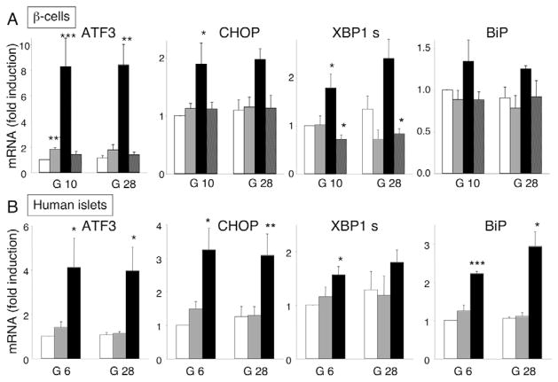
Fig. 5.
FFA activate ER stress signaling in rat primary β-cells and human islets. (A) FACS-purified rat β-cells were cultured for 24 hours in the presence of oleate (gray bars), palmitate (black bars) or oleate plus palmitate (hatched bars) at glucose concentrations of 10 and 28 mM. (B) Human islets were cultured for 48 hours in the absence (white bars) or presence of oleate (gray bars) or palmitate (black bars) at glucose concentrations of 6.1 and 28 mM. ATF3, CHOP, XBP1s and BiP mRNA expression was analyzed by real-time PCR, normalized for the expression level of the housekeeping genes GAPDH (for rat β-cells) or β-actin (for human islets) and expressed as fold induction of the control. The results represent means ± s.e.m. of three (human) or four to seven (rat) independent experiments. *P<0.05, **P<0.01, ***P<0.001 vs control (white bars).

