
Figure 1.
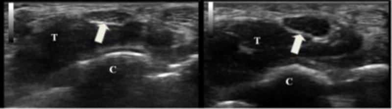
On the left is a cross-sectional view of the median nerve (arrow) at the distal wrist crease in a healthy volunteer. The cross-sectional area of the nerve is 9 mm2 and there is normal nerve echogenicity. On the right is the same view from an individual with carpal tunnel syndrome. The nerve is 19 mm2 and very hypoechoic. T = flexor tendons and C = carpal bones.