
Figure 8. IDE protein expression in breast (upper panel) and ovarian cancer (lower panel) tissue specimens applying different antibodies to IDE.
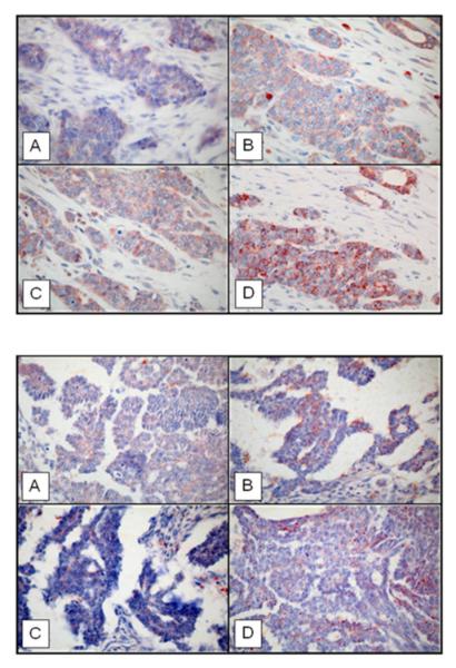
(Magnification x 400). A) rabbit antibody UCG 43/6, University of Chicago, B) mouse antibody #MMS-282R, Covance, C) rabbit antibody #PRB-282C, Covance, and D) rabbit antibody to IDE-peptide p15, Pineda.