Fig. 5.
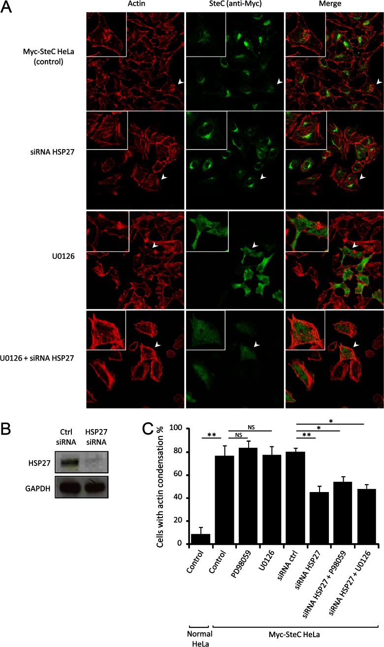
SteC manipulates actin through HSP27. A, representative confocal images of HeLa cells stably expressing Myc-tagged SteC. Cells were immunolabeled with anti-Myc antibody (green), and F-actin was visualized by phalloidin conjugated to Alexa Fluor 568 (red). Cells were treated with siRNA (1 nm control siRNA and 1 nm HSP27 siRNA) and MEK inhibitors (30 μm PD98056 and 10 μm U0126). B, immunoblot analyses of Myc-tagged SteC HeLa in siRNA-transfected cell lysates. Immunoblots detected by anti-HSP27 antibody (top panel) and anti-GAPDH antibody (control (Ctrl), bottom panel) are shown to confirm knockdown of HSP27. C, quantification of cells with actin condensation. The means (±S.D.) of proportion of cells with actin condensation were determined from at least three independent experiments, in which a total of more than 100 cells were examined. Statistical analyses were performed with two-tailed unpaired Student's t test. *, p < 0.001; **, p < 0.0001.

