Fig. 5.
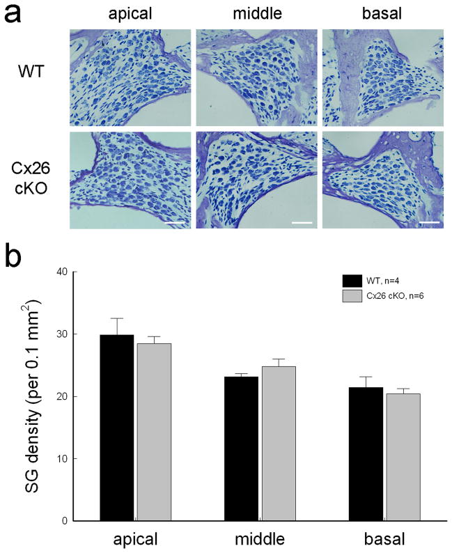
No spiral ganglion (SG) neuron degeneration in Cx26 cKO mice. a: SGs in Rosenthal’s canal at the apical, middle, and basal turn. The sections were stained with toluidin blue. WT littermates served as control. The mice were P30–60 old. Scale bars = 30 μm. b: The density of SGs in Rosenthal’s canal at the apical, middle, and basal turns. There is no significant difference in SG densities between Cx26 cKO mice and WT mice (P > 0.05, one-way ANOVA). Data are expressed as mean ± S.D.

