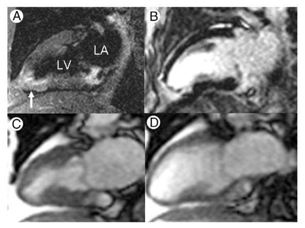
Figure 1. Myocardial Edema at Initial Presentation With NSTE-ACS.
Magnetic resonance images obtained in a 63-year-old female nonsmoker with chest pain, nonspecific electrocardiographic abnormalities, and troponin-I that increased from 0.04 to 2.36 mg/dl over the initial hours of hospital stay. T2-weighted imaging (A; vertical long-axis plane) shows infero-apical edema (arrow), and late postgadolinium enhancement (B) indicates irreversible injury. There is corresponding wall motion abnormality indicated by abnormal myocardial thickening at end-systole (C) compared with end-diastole (D) of a vertical long-axis cine. NSTE-ACS = non–ST-segment elevation acute coronary syndrome.

