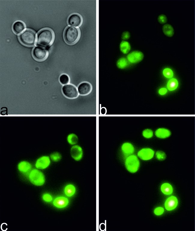
Fig 6.
PDI effect on C. albicans cells during illumination. Cells were incubated in the dark with IA C1330 (100 μM at 37°C). After this, cells were observed under a fluorescence microscope for 30 min. At particular times, pictures were taken at time zero, with white light, directly after incubation in the dark (a) and 5 min (b), 15 min (c), and 20 min (d) after illumination. Panels b to d represent IA fluorescence (excitation wavelength of 360 to 370 nm and emission wavelength of >420 nm).

