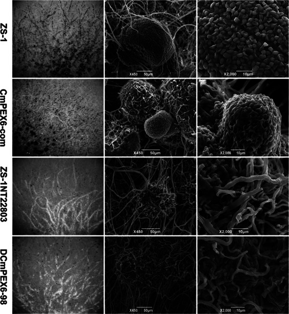
Fig 2.

Microscopic observation of colony morphology and pycnidia formed by nonmutants first and then mutants ZS-1TN22803, DCmPEX6-98, CmPEX6-com, and ZS-1 with SEM. All strains were cultured at 20°C on PDA for 7 days. Column I shows the colony morphology. Column II shows pycnidia near the margin of the colony. Column III shows the conidia formed in the pycnidia. There were no pycnidia or conidia formed in colonies of ZS-1TN22803 or DCmPEX6-98.
