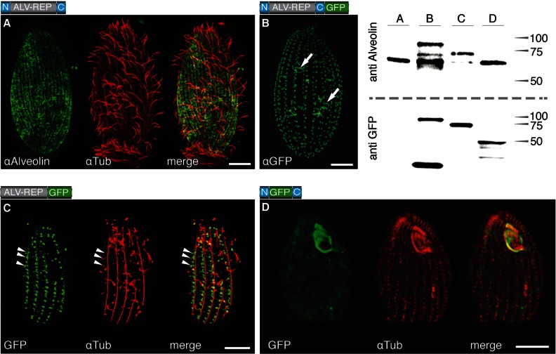
Fig 1.
Localization of TtALV2 and fragments thereof. (A) Endogenous TtALV2 (68 kDa) localizes to patches between the longitudinal microtubules and along the entire inner cell surface. αAlveolin, anti-alveolin; αTub, anti-tubulin. (B) In contrast, the TtALV2::GFP fusion protein (94 kDa) localizes predominantly to the basal bodies and forms fiber-like structures inside the cytosol (arrows). Only in this case was a GFP antibody (αGFP) used; in all other cases, autofluorescence of GFP is shown. (C) Repeat motifs alone fused to GFP (ALV-REP::GFP; 75 kDa) associate with microtubular structures, including transverse microtubules (arrowheads) and the basal bodies. (D) Replacement of the repeat motifs with GFP (N::GFP::C; 46 kDa) targets the fusion protein predominantly to the oral apparatus. The schematics of the constructs are shown at the top left corner of each panel, where N and C represent the nonrepetitive N- and C-terminal domains of TtALV2, respectively. Scale bars, 10 μm. The corresponding Western blots of the protein extracts of the individual strains are shown at the top right. Markers are in kilodaltons.

