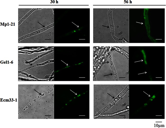
Fig 6.
Distribution of chimeric GFP-Mp1, GFP-Gel1, and GFP-Ecm33. After cultivation at 37°C for 24 or 56 h, the mycelia were harvested, treated with 0.5 M sorbitol, and then analyzed under fluorescent light, using a Zeiss microscope equipped with a 460- to 480-nm excitation filter set, captured with a CCD camera, and edited with the image analyzer program Image (AxioVision Rel.4.6). The cell wall is marked with an arrow.

