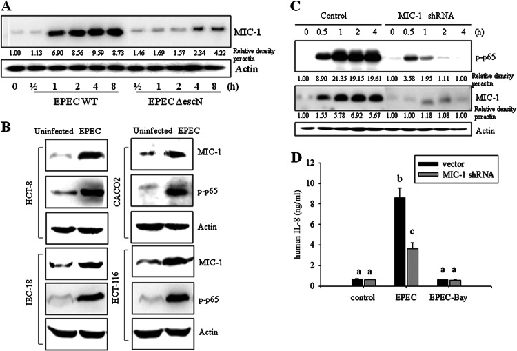
Fig 5.
Roles of MIC-1 in EPEC infection in intestinal epithelial cells. (A) HCT-8 intestinal epithelial cells were infected with wild-type EPEC or EPEC ΔescN at a ratio of 50:1 (bacteria to cells) at each time point. Total proteins from the epithelial cellular lysate were subjected to Western blot analysis. (B) Intestinal epithelial cells (HCT-8, HCT-116, CACO-2, and IEC-18) were infected with EPEC at a ratio of 50:1 (bacteria to cells) for 4 h. Total proteins from epithelial cellular lysates were subjected to Western blot analysis. All results are representative of three independent experiments. (C) Stable cell lines (control and HCT-8 cells stably expressing MIC-1-specific shRNA) were infected with EPEC at a ratio of 50:1 (bacteria to cells) for the indicated times. Total proteins from the epithelial cellular lysate were subjected to Western blot analysis. (D) Stable cell lines (control and HCT-8 cells expressing MIC-1-specific shRNA) were pretreated with vehicle (dimethyl sulfoxide) or 20 μM BAY 11–7082 (an NF-κB inhibitor) and then infected with EPEC for 1 h. IL-8 secreted into the culture supernatant was analyzed using an ELISA. Values are the means ± standard errors of the means (n = 6 to 12). Bars with different letters are significantly different (P < 0.05). Pairwise comparisons were performed using SNK post hoc ANOVA.

